医影在线

标题: CL0109:【】肺泡癌 [打印本页]

作者: 专党zey    时间: 2004-12-26 02:12
标题: CL0109:【】肺泡癌


[本贴已被 翁志蓬 于 2008-9-30 0:49:49 修改过]

作者: 专党zey    时间: 2004-12-26 02:18
标题: 回复:肺泡癌

作者: zhe55555    时间: 2004-12-26 03:11
diagnosis: bac
(bronchioloalveolar carcinoma)

  radiologic findings

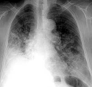
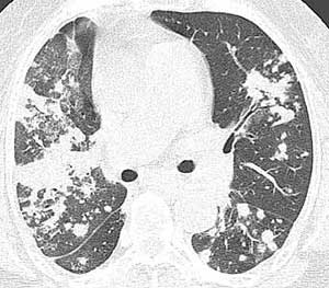
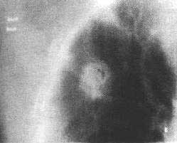
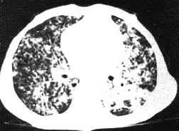
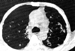
ct scans shows multifocal patchy areas of confluent airspace consolidation and ground-glass attenuation in both lungs.
air bronchograms are well visualized within the consolidations.
some patchy nodules seem to be centrilobular in location.

diffuse form of bac (bronchioloalveolar carcinoma), malignant lymphoma, and active pulmonary tuberculosis can be considered in the pictures like these.


  brief review

  bronchioloalveolar carcinoma is characterized pathologically by lepidic growth with preservation of lung architecture. the most frequent symptoms and signs in patients with bronchioloalveolar carcinoma include cough, sputum, shortness of breath, weight loss, hemoptysis, and fever. bronchorrhea, once considered the clinical hallmark of this disease, is unusual and is a late manifestation seen only with diffuse bronchioloalveolar carcinoma (1, 2).

  the radiologic manifestations of bronchioloalveolar carcinoma are diverse and include single or multiple pulmonary nodules, segmental or lobar consolidation and diffuse airspace disease. alveolar filling disorders are usually seen in forms of segmental or lobar and diffuse airspace disease. the lobar consolidative form accounts for approximately 30% of all bronchioloalveolar carcinomas and corresponds to a mucinous histologic type. the airspace consolidation is caused by growth along the alveolar wall combined with secretion of mucin.

  production of copious amounts of mucin may result in expansion of the involved lobe, leading to bulging of interlobar fissures. the ct angiogram sign is caused by the homogeneous low attenuation of consolidation, which allows vessels to be clearly seen, particularly after intravenous administration of contrast material. the ct angiogram sign is nonspecific and may also be seen in lobar pneumonia, pulmonary lymphoma, extrinsic lipid pneumonia, pulmonary infarction, and pulmonary edema. according to recent report by akira et al (3), diffuse bronchioloalveolar carcinoma can be classified into three patterns: ground-glass opacity, consolidation, and multiple nodules (the most common pattern is ground-glass opacity). areas of ground-glass opacity are found both around and remote from the consolidation or nodule. the involved bronchi in lobar bronchioloalveolar carcinoma show stretching, spreading, and uniform narrowing.


  references

1. clayton f: bronchioloalveolar carcinomas: cell types, patterns of growth, and prognostic correlates. cancer 57:1555-64,1986

2. manning jt, spjut hj, tschen ja: bronchioloalveolar carcinoma: the significance of two histopathologic types. cancer 54:525-534,1984

3. arika m, atagi s, kawahara m, et al: high-resolution ct findings of diffuse bronchioloalveolar carcinoma in 38 patients. ajr 173:1623-1629, 1999
作者: pp    时间: 2004-12-26 05:45
[emb25][emb7][emb2]
作者: 寻芳者    时间: 2004-12-26 07:08
收到谢谢
作者: 权世辉    时间: 2004-12-26 18:48
[emb7]
作者: x-ray    时间: 2004-12-27 02:41
[emb20]
作者: 大鹏    时间: 2004-12-27 02:55
很好!谢谢!美中不足是片子太少!
作者: 阿圣    时间: 2004-12-27 07:18
[emb21]
作者: 黄恩辉    时间: 2004-12-27 19:05
[emb7]
作者: zhouyong0318    时间: 2004-12-27 20:28
[emb6][emb6]
作者: 专党zey    时间: 2004-12-29 16:10
一、bac的组织学分型----看看他的组织分型,跟影像表现关系密切。


  bac在镜下观察其基本特征和4项诊断标准(周围型病灶、非来源于中央支气管、肿瘤细胞沿肺泡间隔生长、肺外无原发性腺癌)已被广大病理工作者接受。有关亚型分类尚无统一意见。各家从自己的认识提出各自的组织学亚型,如manning等将bac分为ⅰ型和ⅱ型两个亚型。阎培莎、李维华根据组织结构和细胞形态将bac分为4型即肺泡型、乳头状型、粘液型和混合型。近期的文献将bac分为3型[2]:粘液型、非粘液型和硬化型。


  1.粘液型:约占bac的42%,形态改变与maning等的ⅰ型bac和阎培莎、李维华[3]的粘液型基本相同。细胞呈杯状细胞样,光镜、电镜和组织化学检查均具粘液细胞的特征,往往无间质纤维化,间质无慢性炎症细胞浸润,形态学上很难与转移性腺癌,特别是来自结肠和卵巢的腺癌鉴别。


  2.非粘液型:约占48%,与manning等提出的ⅱ型bac基本相同。细胞呈立方形或柱状,表面拱出,可有多核巨细胞,嗜酸性核包涵体。间质常有一定程度的结缔组织增生,淋巴细胞浸润;电镜观察见瘤细胞有clara细胞或ⅱ型肺泡上皮细胞分化的特点。ⅱ型肺泡上皮细胞分化的bac可出现核内包涵体,其性质未确定,一般认为是表面活性物质阿朴蛋白(surfactant apoprotein)。


  3.硬化型:约占10%。光镜、电镜下观察均与非粘液型相同。不同之处在于本型肿瘤中央区有硬化灶。有作者将此型归类于“疤痕”,是疤痕诱发癌,还是癌引起的结缔组织增生反应导致硬化?尚不清楚,有实验证明肿瘤内的胶原是新生的,支持疤痕是结果而不是原因。以上3型以非粘液型预后较好。


  从组织学分型可以看出,bac实际上是一类异源性肿瘤,可能来源于细支气管上皮的clara细胞、化生的粘液细胞或ⅱ型肺泡上皮细胞。有人认为来源于支气管上皮的干细胞,向不同方向分化从而显现不同的表型。
作者: liyur17135    时间: 2004-12-31 06:15
[emb1]
作者: 向医生    时间: 2005-1-3 23:35
细支气管肺泡癌(alveolar cell carcinoma)         

[病因病理]
    细支气管肺泡癌系起自于呼吸性细支气管及肺泡上皮的肿瘤。
[临床表现]
    可有咳嗽、咯血丝痰等表现。
[影像学表现]  
    1.x线表现:基本上可见三种类型。
    ①单发结节,多难与周围型肺癌区别。体层或ct检查发现“空泡征”,有诊断价值。
    ②多发结节(广泛型),其结节多较粟粒结节为大,分布可不均匀。往往早期起于肺的一部分,后逐渐蔓延至全肺。
    ③渗出性病变,单一照片确诊较难。上肺野者往往误诊为肺结核;下肺野者往往误诊为肺炎。但抗炎或抗痨治疗均不能控制临床症状的发展及x线病灶的进展。
    2.ct表现:
    ①多灶型表现两肺、一侧肺或某一叶多发病灶,大小不一,呈弥漫结节、团块状或片状影。
    ②单发结节型,常位于肺外围或胸膜下,呈结节状或不规则形,其内有“含气细支气管征”或“空泡征”,病变周边可有“胸膜皱缩征”。
    ③病变也可累及整个叶、段。其内有多发大小不等含气空腔;也可呈均一实变,内有“含气支气管征”,管壁不规整,僵硬和扭曲。
    ④以上各型均可合并有肺门和纵隔淋巴结增大。
[鉴别诊断]
    单发结节型细支气管肺泡癌诊断困难,难与其他孤立性肺结节病变如结核瘤和其他良性肺肿瘤鉴别;多发结节型应与其他多发结节性病变如尘肺、血行播散性肺结核鉴别,痰检和临床症状有助鉴别。当病变局限某一肺叶或肺段时,如有多发空腔,需与纤维空洞型肺结核鉴别。若为均一实变,应鉴别病变包括肺炎,肺梗塞等。
作者: 向医生    时间: 2005-1-3 23:42
标题: 肺泡癌——结节型


细支气管肺泡癌(结节型)。胸片(上图)示右上肺第二前肋间病变阴影淡,轮廓模糊。体层摄影(下图)示肿块边缘呈细小毛刺,密度不均,其间见多个小的透亮区。病理证实为细支气管肺泡癌,肿瘤内部都为结节状的癌组织聚集而成。
作者: 向医生    时间: 2005-1-3 23:47
标题: 肺泡癌——广泛型

细支气管肺泡癌(广泛型)。胸片示两肺广泛分布的粟粒性结节阴影,右肺病变较为密集。


细支气管肺泡癌(广泛型)。ct平扫肺窗(上图)示两肺弥漫分布结节状及斑片状病灶。纵隔窗(下图)示主-肺动脉窗部淋巴结增大,部分肺结节内含有小的空腔。痰检病理证实为细支气管肺泡癌。
作者: zhanyong    时间: 2005-1-4 08:31
呵呵。学习!
作者: xizhwa    时间: 2005-1-5 02:47
[emb25][emb25]
作者: 晨雨    时间: 2005-1-7 04:51
谢谢![emb7]
作者: anxiangyingxiu    时间: 2005-1-10 23:24
有什么临床症状?有没有大量的泡沫痰?
作者: daqingmr    时间: 2005-2-25 21:31
[emb20]
作者: qinpeng    时间: 2005-3-9 08:28
[emb25][emb26][em4]
作者: 冷开水    时间: 2006-2-2 05:12
英文水平有限,请用中文讲解
作者: 寻芳者    时间: 2006-2-24 15:15
好例!!!
作者: ziputao52    时间: 2006-5-8 01:01
和一 些特殊的感染怎么鉴别呢?
作者: drzhang8888    时间: 2006-8-17 08:47
谢谢
作者: jinguoji    时间: 2006-11-6 05:05
学习!
作者: 晨雨    时间: 2008-10-30 19:06
谢谢
作者: 513ytb    时间: 2009-7-8 05:38
肺泡癌 .
作者: fanxinyu    时间: 2010-5-13 02:13
学习!
作者: fanxinyu    时间: 2010-5-26 06:07
学习了
作者: xuqiuyan    时间: 2010-11-13 21:21
肺泡癌的表现真是多种多样,弥漫性的应注意与炎症、结核及其他特殊感染鉴别,紧密结合临床,应想到本病。
作者: shibing    时间: 2012-5-26 15:50
肺泡癌


欢迎光临 医影在线 (http://bbs.radida.com/bbs/) Powered by Discuz! X3.2