医影在线
标题: X0016:[转帖]Chest Radiography and Pulmonary Embolus [打印本页]
作者: rjbjl 时间: 2004-4-24 00:26
标题: X0016:[转帖]Chest Radiography and Pulmonary Embolus
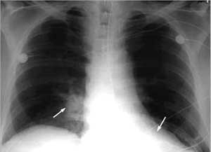
atelectasis and parenchymal densities are quite common.
the areas of atelectasis are more common
in the lower lobe as are the areas of parenchymal density.
[本贴已被 翁志蓬 于 2004-4-23 16:45:57 修改过]
[本贴已被 rjbjl 于 2004-4-23 17:06:03 修改过]
作者: rjbjl 时间: 2004-4-24 00:32
标题: 回复:[转帖]chest radiography and pulmonary embolus
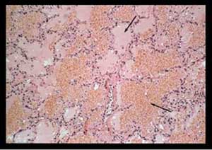
most of these densities are caused by pulmonary
hemorrhage and edema and can be confused
with infectious infiltrates or malignant masses.
[本贴已被 rjbjl 于 2004-4-23 17:05:33 修改过]
作者: rjbjl 时间: 2004-4-24 00:35
标题: 回复:[转帖]chest radiography and pulmonary embolus
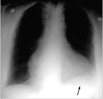
pleural effusions are common and most often
unilateral despite the fact that most clots are bilateral.
these effusions are usually visible when the
patient seeks medical attention.
they are almost always small,
occupying less than 15% of a hemithorax and
rarely increase in size after 3 days. any increase
in size after 3 or 4 days should raise the suspicion
of a pulmonary infection or re-embolization.
作者: rjbjl 时间: 2004-4-24 00:38
标题: 回复:[转帖]chest radiography and pulmonary embolus
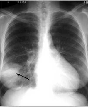
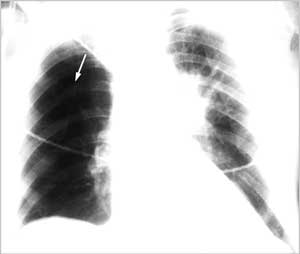
pleural based opacities with convex medial margins
are also known as a hampton';s hump.
this may be an indication of lung infarction. however,
that rate of resolution of these densities is
the best way to judge if lung tissue has been infarcted.
areas of pulmonary hemorrhage and edema resolve
in a few days to one week. the density caused
by an area of infarcted lung will decrease slowly over
a few weeks to months and may leave a linear scar.

作者: rjbjl 时间: 2004-4-24 00:41
标题: 回复:[转帖]chest radiography and pulmonary embolus
a diaphragm may be elevated,
reflecting volume loss in the affected lung.
作者: rjbjl 时间: 2004-4-24 00:46
标题: 回复:[转帖]chest radiography and pulmonary embolus
the central pulmonary arteries may be prominent
either from pulmonary hypertension or the
presence of clot in those arteries.

作者: rjbjl 时间: 2004-4-24 01:02
标题: 回复:x016
a westermark';s sign implies an area of decreased vascularity and
perfusion accompanied by an enlarged central pulmonary
artery on the affected side.
作者: rjbjl 时间: 2004-4-24 01:03
标题: 回复:chest radiography and pulmonary embolus
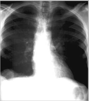
cardiomegally is a non-specific finding but may imply an
enlarged right ventricle as seen in the patient
who presented with large bilateral pulmonary emboli.

作者: rjbjl 时间: 2004-4-24 01:04
标题: 回复:chest radiography and pulmonary embolus
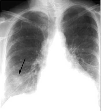
pulmonary edema is an uncommon finding in this group.
this patient was originally diagnosed as suffering
from pulmonary edema but in fact had multiple areas of infarction.

in conclusion, the chest x-ray can be normal
in a minority of cases and abnormalities,
when present, are often non-specific.
作者: rjbjl 时间: 2004-4-24 01:07
终于发完了!/高兴/高兴/大笑
作者: lkc8963 时间: 2004-4-26 06:47
楼上朋友真是太厉害了,看了好久连中西合壁功夫都使出来了也只看了个大概,提个建议--这样也许不太适用于普及和提高,您说对吗?
作者: rjbjl 时间: 2004-4-26 07:00
?
作者: jiajie 时间: 2004-4-28 15:29
片子的确好
用中文会更好
作者: yangxuedong 时间: 2004-5-1 03:12
不错的介绍
作者: liyj000 时间: 2004-5-2 05:21
rjbjlj朋友,你将x线征象与发病机理相结合一起陈述,是迫使我们学英语。谢谢啊
大部分看的懂
作者: 潘建华 时间: 2004-6-2 04:40
要想发展就要学外语,可惜我年龄较大,这方面不可能有多大发展了,建议用中文,不好意思。
作者: guduchunmeng 时间: 2004-6-2 06:09
[emb18]认识abc是在学数学是学的。现在再学英语?[emb11]打不死是不学了。
作者: jinguoji 时间: 2006-11-6 07:42
不好意思,英文基础太差。
作者: shibing 时间: 2012-9-1 14:17
不好意思,英文基础太差。
作者: lyming00 时间: 2012-9-20 09:28
不好意思,英文基础太差。
作者: shibing 时间: 2014-12-18 09:54
ct
| 欢迎光临 医影在线 (http://bbs.radida.com/bbs/) |
Powered by Discuz! X3.2 |